Scientists unlock secrets of brain
Yale researchers probe mental activity taking place as two people engage in conversation
To the untrained eye, the graph looked like a volatile day on Wall Street, with jagged peaks and valleys, but it was not describing economics. It was a glimpse into the brains of Shaul Yahil and Shaw Bronner, two researchers at a Yale University lab, as they had a chat.
"This is a fork," Yahil said, describing the image on his computer. "A fork is something you use to stab food while you're eating it. Common piece of cutlery in the West."
"It doesn't look like a real fancy sterling silver fork, but very useful," Bronner replied.
The jagged, multicolored images showed two brains in conversation. The brain-tracking technology at work is just a small part of the quest to answer abiding questions about the workings of a 1.4-kilogram piece of fatty tissue containing nearly 100 billion densely packed nerve cells, acting through circuits with maybe 100 trillion connections. How does it let us think, feel, act and perceive our world? How does this complex machine go wrong and make people depressed or delusional or demented?
Such questions spurred President Barack Obama to launch the BRAIN initiative in 2013 to spur development of new investigative tools. Europe and Japan are also pursuing major efforts in brain research.
With a collection of sophisticated devices, scientists are peering inside people's brains for clues about what makes us tick.
At the Yale lab, Yahil and Bronner were demonstrating a technique to investigate how our brains let us engage with other people.
That's one of the most basic questions in neuroscience, said lab director Joy Hirsch.
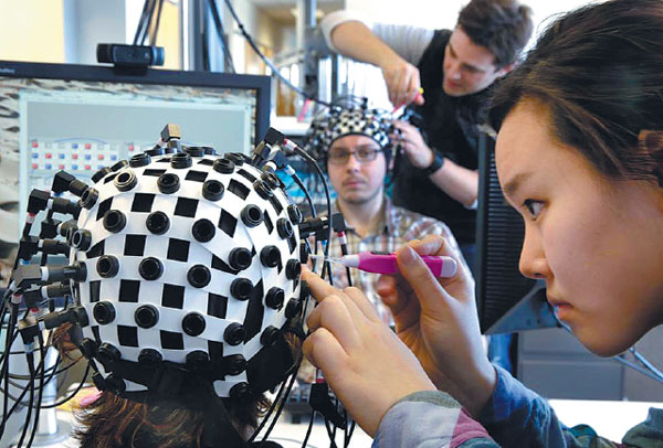
As the two researchers chatted, each wore a black-and-white skullcap from which 64 slender black cables trailed away like dreadlocks.
At the tip of half of those fiber optic cables, weak laser beams slipped through their skulls and penetrated about 2.5 centimeters before bouncing off blood and being reflected back to be picked up by the other half of the cables.
Those reflections reveal how much oxygen that blood was carrying. And since brain circuits use more oxygen when they're busier, the measurements provided an indirect index to patterns of brain activity as Bronner listened to Yahil and replied, and vice versa.
The most widely used brain-mapping technique is called functional magnetic resonance imaging, or fMRI, which uses oxygen levels in blood as tracers of brain cell activity. It penetrates deeper into the brain, using powerful magnetic fields to seek subtle signals and track blood oxygen levels on a tiny scale.
The fMRI technology can detect small changes in brain activity that are associated with tackling particular tasks. And it can show the activity of a brain that is not focused on doing a task.
Scientists are studying what this resting state, when the brain continues to hum along, can reveal about it and its illnesses.
Another major emphasis in brain mapping these days is delineating the circuitry that lets the brain operate.
Some brain-scanning research rises from the informative to the truly startling, like decoding - looking at brain activity patterns to figure out what somebody is seeing, or even thinking about.
In 2011, for example, researchers reported that they could reconstruct very rough visual replicas of movie clips that people were watching while their brains were scanned. And two years later, Japanese scientists reported evidence that they could get some idea of what people were dreaming about.
In the near term, decoding technology might help people whose medical condition prevents normal conversation, said Jack Gallant of the University of California, Berkeley.
|
Undergraduate students attach laser probes to brain activity subjects at the Yale Brain Function Lab in New Haven, Connecticut, during a demonstration of brain mapping technology. At one end of each of the 64 fiber-optic cables in each headpiece, weak laser beams see about an inch into the subjects' brains to detect blood flow. Richard Drew / AP |


