Science and Health
A visual odyssey through the secrets of the brain
By Abigail Zuger, M.D. (China Daily)
Updated: 2010-12-19 08:48
 |
Large Medium Small |
Who has seen the mind? Neither you nor I - nor any of the legions of neuroscientists bent on opening the secrets of that invisible force, as powerful and erratic as the wind.
The experts are definitely getting closer: the last few decades have produced an explosion of new techniques for probing the blobby, unprepossessing brain in search of the thinking, feeling, suffering, scheming mind.
 |
|
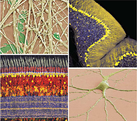
Clockwise from top left: photomicrographs of spiny neuron (2009); cerebellar Purkinje neurons (2003); spiny neuron (2009); chick retina (2008). [Photos by Thomas Deerinck and Mark Ellisman; Aric Agmon; Thomas Deerinck and Mark Ellisman; Andy Fischer] |
But the field remains technologically complicated, out of reach for the average nonscientist, and still defined by research so basic that the human connection, the usual "hook" by which abstruse science captures general interest, is often missing.
Carl Schoonover took this as a challenge. Mr. Schoonover, 27, who is midway through a Ph.D. program in neuroscience at Columbia University in New York, decided to draw the general reader into his subject with the sheer beauty of its images.
So he has compiled them into a glossy new art book. "Portraits of the Mind: Visualizing the Brain From Antiquity to the 21st Century," newly published by Abrams, includes short essays by prominent neuroscientists and long captions by Mr. Schoonover - but its words take second place to the gorgeous imagery, from the first delicate depictions of neurons sketched in prim Victorian black and white to the giant Technicolor splashes the same structures make across 21st-century LED screens.
Scientists are often seduced by beauty. Sometimes the thrill is the magic of a fabulous technique for getting at elusive data.
Consider a blurry little black-and-white photograph of a smiley-face icon, so fuzzy and ill-defined it looks like a parody of the Shroud of Turin. The picture is actually a miracle in its own right: the high-speed video camera that was trained on the exposed brain of a monkey staring at a yellow smiley face. As the monkey looked at the face, blood vessels supplying nerve cells in the visual part of the monkey's brain transiently swelled in exactly the same pattern. We can tell what was on the monkey's mind by inspecting its brain. The picture forms a link, primitive but palpable, between corporeal and evanescent, between the body and the spirit. And behind the photo stretches a long history of inspired neuroscientific deductions and equally inspired mistakes, all aiming to illuminate just that link.
It's only fitting that the story should be a visual one, for the visuals had the ancients fooled for millennia.
Aristotle, for example, concluded that the brain's moist coils served only to cool the heart, the obvious home of the rational soul. Only when the long ban on dissection petered out in the Renaissance did the ventricles prove to be empty space.
In 1873 the Italian scientist Camillo Golgi developed a black stain to highlight the micron-thin neural strands. Fifteen years later the Spanish scientist Santiago Ramón y Cajal, deploying the stain with virtuoso dexterity, presented the world with visible populations of individual neurons. The roots, or dendrites, of these elongated nerve cells gather information. The trunks, or axons, transmit it.
Now those same skeletal silhouettes glow plump and brightly colored, courtesy of inserted genes encoding fluorescent molecules. The most dramatic variation on these methods for highlighting neurons in living color, dubbed the Brainbow by its inventors, turns the brains of living mice into wild neon forests of branching trees. The electrochemical circuitry that propels information around that forest, from nerve to nerve, has generated its own fabulous images.
Meanwhile, the traffic in long groups of neurons all coursing together around the brain becomes visible with a variation on the standard scanning technique called diffusion M.R.I. Here the neurons do look just like pasta, slightly beaded, draped and purposeful. But if the structure is destroyed (by a stroke, for instance) the strands shatter into fragments, the information highway broken.
In the book's final essay, Joy Hirsch, a neuroimaging specialist at Columbia, sympathizes with readers who hate the idea that they - their essential selves, their likes and dislikes, their premonitions, biases and life decisions - are nothing but neural circuits.
"These cells and molecules, awash in various neurochemical cocktails in my basal ganglia, are presumably the basis for my love and attachment to my husband," she writes. "Earlier in my academic journey I would have resisted this unavoidable fact of biology on the misguided rounds that a physical basis would diminish the grandeur and centrality of my choice of a life partner."
Now, Dr. Hirsch says she joyfully embraces "the astonishing unity of the physical brain and the mind." And she doesn't see that anyone has much choice about accepting it.
"People assumed for thousands of years that there must be something else," the science writer Jonah Lehrer writes in the introduction. "And yet, there is nothing else: this is all we are."
The New York Times